SR-B1 and PGRMC1 Expression in Canine Uterine Macrophages
Cordula Gabriel
DOI10.21767/2476-1974.100035
Cordula Gabriel*
Department of Pathobiology, Institute of Anatomy, Histology and Embryology, University of Veterinary Medicine, Vienna, Austria
- *Corresponding Author:
- Cordula Gabriel
Department of Pathobiology, Institute of Anatomy
Histology and Embryology, University of Veterinary Medicine
Vienna, Austria
Tel: +431250773403
Fax: +431250773490
E-mail: Cordula.Gabriel@vetmeduni.ac.at
Received date: July 03, 2017; Accepted date: July 05, 2017; Published date: July 10, 2017
Citation: Gabriel C (2017) SR-B1 and PGRMC1 Expression in Canine Uterine Macrophages. Reproductive Immunol Open Acc Vol.2 No.3:35. doi: 10.21767/2476-1974.100035
Copyright: © 2017 Gabriel C. This is an open-access article distributed under the terms of the Creative Commons Attribution License, which permits unrestricted use, distribution, and reproduction in any medium, provided the original author and source are credited.
Image Article
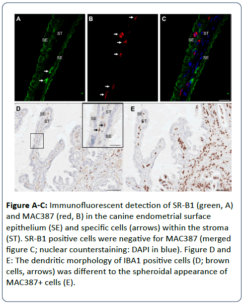
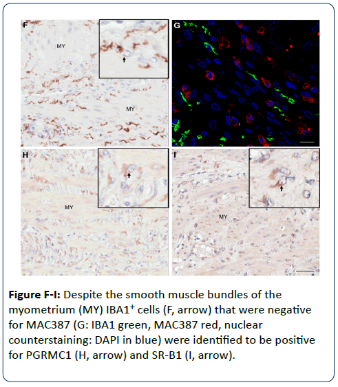
In a previous study [1] the scavenger receptor SR-B1 was identified to be upregulated in the progesterone responsive metestrous and pyometra affected canine uterus. In a follow-up study SR-B1+ expression was identified in IBA1+/MAC387- stromal cells, which were therefore characterized as macrophages (Figures A-E). A comparable cell population was identified between myometrial smooth muscle bundles (Figures F-I). These SR-B1+/IBA1+ cells were positive for PGRMC1 (Figure H). Dressing et al. [2] summarized data suggesting that progesterone acts upon immune cells via non-genomic pathways rather than nuclear progesterone receptors, but data are inconsistent. Our preliminary findings strengthen the hypothesis that these PGRMC1+/SR-B1+/IBA1+ macrophage-like cells might be activated or recruited via progesterone in the canine uterus.
Figure A-C: Immunofluorescent detection of SR-B1 (green, A) and MAC387 (red, B) in the canine endometrial surface epithelium (SE) and specific cells (arrows) within the stroma (ST). SR-B1 positive cells were negative for MAC387 (merged figure C; nuclear counterstaining: DAPI in blue). Figure D and E: The dendritic morphology of IBA1 positive cells (D; brown cells, arrows) was different to the spheroidal appearance of MAC387+ cells (E).
References
- Gabriel C, Becher-Deichsel A, Hlavaty J, Mair G, Walter I (2016) The physiological expression of scavenger receptor SR-B1 in canine endometrial and placental epithelial cells and its potential involvement in pathogenesis of pyometra. Theriogenology 85: 1599-1609.
- Dressing GE, Alyea R, Pang Y, Thomas P (2012) Membrane progesterone receptors (mPRs) mediate progestin induced antimorbidity in breast cancer cells and are expressed in human breast tumors. Horm Cancer 3: 101-112.
Open Access Journals
- Aquaculture & Veterinary Science
- Chemistry & Chemical Sciences
- Clinical Sciences
- Engineering
- General Science
- Genetics & Molecular Biology
- Health Care & Nursing
- Immunology & Microbiology
- Materials Science
- Mathematics & Physics
- Medical Sciences
- Neurology & Psychiatry
- Oncology & Cancer Science
- Pharmaceutical Sciences


