Mechanism Of Cytomegalovirus Penetration Through Erythrocyte Membrane
Lucenko Michael T and Andrievskaya Irina A
DOI10.21767/2476-1974.100028
Lucenko Michael T* and Andrievskaya Irina A
Federal State Budgetary Scientific Institution “Far Eastern Scientific Center of Physiology and Pathology of Respiration”, Blagoveshchensk, Russian Federation
- *Corresponding Author:
- Lucenko Michael T
Federal State Budgetary Scientific Institution “Far Eastern Scientific Center of Physiology and Pathology of Respiration”
Blagoveshchensk, Russian Federation
Tel: +79143929709
E-mail: lucenkomt@mail.ru
Received Date: January 17, 2017; Accepted Date: March 20, 2017; Published Date: March 24, 2017
Citation: Michael LT, Irina AA (2017) Mechanism of Cytomegalovirus Penetration through Erythrocyte Membrane. Reproductive Immunol Open Acc 2:28. doi: 10.4172/2476-1974.100028
Copyright: © 2017 Michael LT, et al. This is an open-access article distributed under the terms of the Creative Commons Attribution License, which permits unrestricted use, distribution, and reproduction in any medium, provided the original author and source are credited.
Abstract
Objective: To study the effect of cytomegalovirus infection on histidine concentration in erythrocytes of peripheral blood in pregnant women at 27-28th week of gestation and virus penetration through the erythrocyte membrane and to demonstrate the morphofunctional alterations of the latter. The objective of this study is to demonstrate the mechanism of cytomegalovirus penetration through the erythrocyte membrane as well as to describe the process of its structure damage which leads to the accumulation in the peripheral blood of degenerative forms of erythrocytes that lose their ability to deformability. This creates the conditions for the formation of hemic anaemia in a pregnant woman. The research has been conducted on the basis of current data and our own studies.
Materials and methods: 75 pregnant women at 27-28th weeks of gestation were examined, including 55 CMV-seropositive ones with CMV-infection exacerbation at 27th week of gestation. All the studies were carried out at the Federal State Budget Scientific Institution “Far Eastern Scientific Centre of Physiology and Pathology of Respiration” during one year time. Histidine activity in peripheral blood erythrocytes was found according to the method suggested by Lucenko Michael T and Andrievskaya Irina A.; fatty acid peroxides in erythrocyte membrane-by Winkler-Schultze method; erythrocyte membrane microviscosity was found by spectrofluorimetric method of lateral diffusion of pyrene. Erythrocyte deformability was also found in accordance with the method of Lucenko Michael T and Andrievskaya Irina A. The calculation of deformed erythrocytes was done using automated cytophotometric device “Mekos” (Russia). Erythrocyte membrane condensation was measured in pixels by means of cytophotometric camera “Pixera” (USA).
Results: During the exacerbation of CMV infection at 27-28th weeks of gestation the amount of distal histidine was lower 2.13 times per one erythrocyte. The level of unsaturated fatty acids increased 2.9 times, of fatty acid peroxides in erythrocyte membranes increased 3.3 times, which resulted in 1.7 times increase in erythrocyte membrane microviscosity in the lipid bilayer and in 6.8 times increase in erythrocyte deformability.
Conclusion: The exacerbation of chronic CMV infection at 27-28th weeks of gestation causes the virus intrusion into erythrocyte membrane damaging the distal histidine content, which promotes further penetration of proteins of viral envelope and tegument into the erythrocyte. That results in the disturbance of phospholipids bond with erythrocyte membrane proteins, increase in fatty acid peroxides in erythrocytes and decrease in the membrane microviscosity. This promotes the accumulation of deformable erythrocytes in peripheral blood thus causing the threat of anemia formation in pregnant woman at late gestation.
Keywords
Chronic cytomegalovirus infection; Pregnancy; Erythrocytes; Histidine.
Background
It is known that the exacerbation of CMV infection during pregnancy provokes the spread of the virus throughout the body and tissue infection. Long-term persistence of CMV in the body is accompanied by involving immune competent and hemopoetic cells into the pathological process. Erythrocytes participate actively in the process of interaction with the virus implemented through the glycophorin receptors. This results in the initiation of proteolytic and hydrolytic processes that alter the redox state and energetic balance of the cell and inhibit enzymatic reaction. As studies have shown, erythrocyte metabolism disturbance begins after the direct contact of erythrocyte membrane receptors with the cytomegalovirus envelope containing gp64 protein and involving in this process a distal portion of histidine [1-6]. The decrease in proteins with histidine leads to pH lowering to 5 [6,7] and promotes a further penetration of virus proteins, especially of tegument proteins, into the erythrocyte. This has a damaging effect on membrane proteins with the manifestation of destructive phenomena in phospholipid layer and an abrupt increase in erythrocyte membrane microviscosity, especially of H152, H156. Owing to gp64 protein there is a rapid penetration of the virus into erythrocyte. Histidine with clusters H152, H155, H156 is hydrogen donors and triggers for these processes initiation [8].
Methods
Research design
A prospective case-control study was done.
Compliance criteria
Study inclusion criteria were exacerbation of CMV-infection at 27-28th week of gestation with the clinical features of acute respiratory viral infection and persistent clinical remission of herpes-virus infection.
Study exclusion criteria were primary CMV-infection, exacerbation of other inflammatory diseases of extragenital pathology and presence of sexually transmitted infections. The primary CMV-infection was clinically diagnosed by the presence in the peripheral blood of class M antibodies to CMV (immunoglobulin Ig), low avid IgG (avidity index <65%), as well as CMV DNA detected by polymerase chain reaction (PCR) method in blood or urine; chronic CMV infection exacerbation was diagnosed by presence of IgM to CMV, high avid IgG (avidity index >65%), as well as CMV DNA in the scrapes of buccal epithelium and cervix uteri mucosa.
Conditions for the research
All studies (clinical, biochemical, histochemical) were conducted at the Federal State Budget Scientific Institution “Far Eastern Scientific Center of Physiology and Pathology of Respiration” (Blagoveshchensk). No specific factors that might affect the conclusions have been detected.
Research outcome
Histidine activity in peripheral blood erythrocytes, the composition and alterations of phospholipids in erythrocyte membranes, fatty acid peroxides in erythrocyte membranes, erythrocyte membrane microviscosity, erythrocyte deformability disturbance and the increase in number of their degenerative forms in peripheral blood, the decrease in oxyhemoglobin level in pregnant women in the third trimester of gestation were the main assessed results.
Research duration
Material collection was done during a year. Samples of blood serum, urine, buccal epithelium and of the cervical canal content were studied simultaneously following biomaterial sampling.
Description of medical intervention
Blood samples were taken from ulnar vein at 27-28th week of gestation to perform histochemical reactions in erythrocyte membrane and to find out the histidine content in separately taken erythrocytes. There was done PCR analysis, and immuneenzyme studies. Cervical canal content was also collected for PCR-analysis.
Methods for research outcome registration
To perform PCR, blood samples from the patients under survey were put into standard vacuum test-tubes with coagulant in amount of 5 ml. Blood samples not containing coagulant were used for serologic tests. The extraction of mononuclear blood cells for PCR was performed using fikkoll-urograffin solution, density 1.077 g/ml (NPO “DNA-technology”, Russia). Serologic tests were conducted in paired sera with 10-14 days interval. Morning urine for PCR-analysis was taken into the sterile container of 60 ml volume. Samples of buccal epithelium and of cervical canal content were collected into standard plastic test-tubes containing 0.5 ml of saline solution, using sterile cotton swab. Histidine content in peripheral blood erythrocytes was found by histochemical method [9]. The whole blood erythrocytes were incubated using Pauli reagent which consists of the solutions in equal volumes: solution 1 is made of sulfanilic acid (1%) in 1M hydrochloric acid; solution 2 of 5% aqueous solution of sodium nitrite. Before the use both solutions were stored in the fridge at 4°C. The equal volumes of solutions 1 and 2 were mixed (also in the cold) 5 minutes before the use. 500 MKL of prepared Pauli reagent and 200 MKL of the whole blood, taken from the ulnar vein from a person having an empty stomach into the standardized PUTH EDTA k3 vacuum tubes, were mixed with the coagulant. The material was stored for 10 minutes at room temperature; after that monolayer smears were prepared in centrifuge “Diff-Spin-2”. The obtained smears were dried and studied by immersion method with the means of digital microscope MEIJI (Japan) working on the software “Scion” enabling to allocate each separately taken red blood cell on the smear image and to determine automatically the reaction quantity to histidine. The calculations were done per 100 erythrocytes both in the blood from a pregnant woman in latent phase and from women with the exacerbation of CMV-infection.
Morphofunctional forms of erythrocytes in blood smears were calculated at the automated cytophotometric device “Mekos” (registered certification MH RF 29/10010198/1282-01, Russia). ErythroÃÆââ¬ËÃâÃÂyte membrane density was measured using digital camera “Pixera” (USA) by the Bio Vision program at the constant range of 60 pixels, which corresponded to the morphofunctional state of degenerative forms of erythrocytes. Erythrocyte deformability was found by means of cytophotometric device “Mekos”. Erythrocyte membrane microviscosity was measured via spectrofluorimetric method of lateral diffusion of pyrene at the spectrophotometer “Hitachi” (Japan) [10]. Phospholipids level was found by thinlayer chromatography of Kirchner Y [11]. Erythrocyte deformability was studied according to Lucenko Michael T and Andrievskaya Irina A formula [12]. Oxygenated hemoglobin level was measured by Evelini and Melloy method [13]. Fatty acid peroxides in erythrocyte membranes were found by histochemical method [14]. Protein-lipid complexes condensation in the erythrocyte membranes was defined using digital camera “Pixera” by BioVision program (USA) Table 1 [15].
| Reaction | Incubation solution composition | Amount |
|---|---|---|
| Histidine Fatty acid peroxides By Winkler-Schultze method (Pearse E. 1962) |
Solution 1 - sulfanilic acid (1%) Solution 2 - 5% aqueous solution of sodium nitrite 576 mg of α-naphtol(ICN Biomedical,USA) in 6 ml of 1N NaOH (Reakhim,RF) brought to 50 ml with distilled water 691 mg of n-amino-N,N-dimethyl-phenylenediamine (Laverana, Russia) brought to 50ml with distilled water 0.2% Lugol’s solution (iodine) 0.005% solution of lithium carbonate (IcN Biomedical, USA) |
1M in HCl 0.25 ml 0.25 ml 0.5 0.5 |
Table 1: Composition and amount of reagents used for histochemical tests.
Erythrocyte Membrane Microviscosity
It was found out that the disturbance of the lipid bilayer microviscosity occurs during the erythrocyte membrane protein oxidation. Using the most sensitive fluorometer Hitachi (Japan), there was revealed the increase in the structuring of the anular lipid zone and the decrease in the lipid bilayer polarity in the erythrocyte membrane when exposed to cytomegalovirus infection.
Pyrene fluorescence was measured in the medium: NaCl-145 mM, tris-HCl-10 mM (pH 7.4). There was found out the following: pyrene excimerization rate in the area of anular and total lipids (L/L at λÃÆà  Ãââ⢠285 and 35 nm) and the polarity of the environment of pyrene molecules (L/L at λÃÆà  Ãââ⢠285-340 nm). The rate of energy migration from the tryptophan residues of the protein to pyrene was also calculated.
Oxyhemoglobin Determination
Oxyhemoglobin concentration was determined on spectrophotometer StatFa x 2100 (USA). The measurement of absorption spectra was performed at six analytical wave lengths in the range of 450-650 nm in hemolyzed blood solution (1%). The subsequent calculations of oxyhemoglobin concentrations were performed based on the measured optical density by means of software. The method is simple and reliable.
Automated Device “Mekos”
This device is an automated cytophotometric device supplied with the digital camera Digital Colour Video Camera JAC and connected to a computer system. The peripheral blood monolayer smear was prepared by means of centrifuge DiffSpin 2 (USA). In accordance with the researcher’s desire the erythrocyte image on the computer screen can be displayed automatically with the resolution of 100-500-1000 and so forth. Next these erythrocytes are differentiated in a percentage ratio and the findings are reflected in the table. As the second stage we make the gallery of the same type of erythrocytes: discocytes, target-shaped erythrocytes, echinocytes, and degenerative erythrocytes. This gallery can be transferred to another computer in order to study certain erythrocytes through the PhotoShop program. It enables to calculate their size and process them for the membrane density using the digital camera by BioVision program (USA). Erythrocyte density is calculated in pixels.
“Mekos” Program can Provide other Indicators that Characterize the Red Blood Cells:
• Erythrocyte diameter
• Erythrocyte polarization
• The specific optical density
• Erythrocyte volume area (erythrocyte surface area)
Using erythrocyte surface area indicators we were able to calculate the deformability of variously shaped erythrocytes. The formula is recorded in the manuscript. It was approbated in rospatent and the patent #2463597 was received [12].
The ethical review
The whole study was conducted with regard to the requirements of World Medical Association Declaration of Helsinki (2008) and the Rules of Clinical Practice in the Russian Federation adopted by Order of the Ministry of Health of the Russian Federation dated June 19, 2003 #266. The study was conducted within the framework of the SRW 059 and can be considered not contrary to the fundamentals of medical ethics. No further recommendations were given by the Ethics Commission at the Far Eastern Scientific Center of Physiology and Pathology of Respiration (Protocol #113 dated October 6, 2016). The written information consent was obtained from all pregnant women.
Statistical Analysis
Statistical analysis and processing of obtained data presented by two selections were performed by t-test according to IBM SPSS Statistics.
Survey Participants
The study included 75 pregnant women (Russians) at 27-28th week of gestation including 55 CMV-seropositive women with the exacerbation of chronic CMV-infection (main group) and 20 CMV-seronegative women (control group). The average age of pregnant women in the main group was 24.8 ± 0.3 years old and there was no statistically significant difference regarding the average age of pregnant women in control group; they were 23.5 ± 0.5 years old.
Key Findings of the Research
As it was demonstrated by our research, at the exacerbation of cytomegalovirus infection during the CMV contact with the membrane there is the decrease in distal histidine content in peripheral blood erythrocytes till 12.0 ± 0.18 pixels/mmk2 per one hundred counted cellular elements (Figures 1 and 2); control group -36.5 ± 1.2 pixels/mmk2; p,=0.0002.

Figure 1: Reaction to histidine in peripheral blood erythrocytes from pregnant women at 27-28th weeks of gestation. Control group. Magnification 10 x 100.

Figure 2: Reaction to histidine in peripheral blood erythrocytes from pregnant women during the exacerbation of CMV-infection with the antibody titer 1:1600. Magnification 10 x 100.
Gp64 viral fusion protein fixed in its outer envelope penetrates deeper and deeper into the erythrocyte membrane. It causes the destruction of the outer surface area of the erythrocyte and allows gp60 to penetrate deeper into the cellular element. Due to the effect of tegument proteins the toxic influence on enzymatic system and hemoglobin oxygenation is manifested. It is noted that there is the violation of phospholipids bonds with the skeletal proteins, which results in the phospholipids structure alterations: phosphatidylethanolamine concentration decreases to 20.03 ± 0.035% (control-23.95 ± 0.75%; p=0.0020). Lysophosphatidylcholine level increases up to 6.0 ± 0.2%. These alterations promote the increase of unsaturated fatty acids amount in erythrocytes from pregnant women with the exacerbation of CMV-infection up to 62.0 ± 1.3 pixels/mmk2 compared to 21.00 ± 0.4 pixels/mmk2 in control group, p=0.0000. Structural violations in phospholipids induce the increase of microviscosity in the erythrocyte membranes [10]. In the lipid bilayer in seropositive patients it increases up to 0.84 ± 0.008 Fe/Fm (control-0.60 ± 0.005 Fe/Fm; p=0.0023), and in the zone of lipid-protein interactions it grows up to 1.09 ± 0.04 Fe/Fm; in control-0.70 ± 0.06 Fe/Fm, p=0.0003). Applying “BioVision” method which enables to define the structural density at various ranges by means of cytophotometric camera “Pixera” (USA), we have discovered that during the exacerbation of cytomegalovirus infection and virus penetration through the red blood cell membrane it abruptly becomes condensated [15]. Using cytophotometric device “Mekos” (Russia) enabling to analyze automatically the quantitative and qualitative structure of peripheral blood erythrocytes, it was found out that during the exacerbation of CMVI there are up to 13.0 ± 0.09% of degenerative forms of erythrocytes (Figure 3) and up to 8.0 ± 0.7% of echinocytes (Figure 4) in the peripheral blood of pregnant women in the third trimester of gestation. The number of discocytes decreases to 79.0 ± 1.8% (Figure 5 and Table 2).

Figure 3: Degenerative erythrocyte gallery in the peripheral blood from a pregnant woman in the third trimester of gestation after the exacerbation of cytomegalovirus infection to antibody titer to cytomegalovirus 1:1600. At the range 65 pixels/mmk2 erythrocyte membrane is highly condensed. Magnification 10 × 100.

Figure 4: Peripheral blood erythrocytes from a pregnant woman in the third trimester of gestation. Echinocytes gallery. According to BioVision program at the range 65, up to 52 ± 1.2 pixels/mmk2 of condensed sites are visualized. Magnification 10 x 100.

Figure 5: Peripheral blood discocytes from a pregnant woman in the third trimester of gestation without history throughout the gestation. There are solitary sites in the membrane visualized by smear processing according to Bio Vision program at the range 65. The quantity of condensed sites is 15 pixels/mmk2. Magnification 10 x 100.
| Erythrocyte types studied according to Bio Vision program at the identical range-65 pixels using Pixera camera | control | Exacerbation of cytomegalovirus infection up to antibody titer to cytomegalovirus 1: 1600 at 27-28th week of gestation | Statistics |
|---|---|---|---|
| Discocytes | 92.2 ± 2.1 | 79.0 ± 1.7 | p=0.0000 |
| Echinocytes | 3.0 ± 0.2 | 8.0 ± 0.07 | p=-0.003 |
| Degenerates | 5.0 ± 0.4 | 13.0 ± 0.09 | p=0.0004 |
Table 2: Calculation of variously shaped erythrocytes at the latent phase of gestation and during the exacerbation of cytomegalovirus infection in the third trimester of gestation by means of automated cytophotometric device ’’Mekos”.
The violation of membrane protein-lipid bilayer leads to the fatty acid peroxides increase up to 80.35 ± 1.9 pixels/mmk2 (Figure 6) (control-25.2 ± 0.8 pixels/mmk2) (Figure 7). P, = 0.0000

Figure 6: Peripheral blood erythrocytes from a pregnant woman in the third trimester of gestation at the exacerbation of cytomegalovirus infection to antibody titer to cytomegalovirus 1:1600. There is an increase of fatty acid peroxides concentration in erythrocyte membranes up to 80.35 ± 1.9 pixels/mmk2. Winkler-Schultze test for fatty acid peroxides. Magnification 10 ÃÆââ¬ËÃâââ¬Â¦ 100.

Figure 7: Peripheral blood erythrocytes from a pregnant woman not being sick throughout the gestation. Control Fatty acids peroxides concentration in erythrocyte membranes is 25.2 ± 0.8 pixels/mmk2. Winkler-Schultze test for fatty acid peroxides. Magnification 10 ÃÆââ¬ËÃâââ¬Â¦ 90.
The revealed defect in erythrocyte membrane leads to the increase in their degenerative forms in peripheral blood. This drastically reduces their deformability index to 0.132 ± 0.002 conventional units compared to the control-0.88 ± 0.004 conventional units (p,=0.0002). Hypoxia (HbO 96%) developing in pregnant women during the exacerbation of cytomegalovirus infection is confirmed by the increase in hypoxia-inducible factors (Hif’s) up to 4.8 ng/ml (control-2.7 ng/ml; p,=0.0023). Hif-1α and Hif-2β have control over oxygen transfer to the tissues through the regulation of gene product expression involved in the cellular energetic metabolism. Hif-1α associates with von Hippel-Lindau (VHL) gene protein which is capable to have a Hif-1α degradation, form the active complex with β- substance, and to becomes stable, as a result [16,17].
 Fully expanded formula can be expressed as follows:
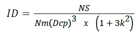 ID-Deformability index,
N-Erythrocyte number under study,
S-One erythrocyte average square in mkm,
V-Erythrocyte volume,
m-standard deviation,
Dcp-Erythrocyte average diameter,
k-Erythrocyte diameter coefficient of variation.
The formula for determining erythrocyte deformability in peripheral blood. The formula was developed by Lucenko Michael T., Andrievskaya Irina A [12].
Discussion
Cytomegalovirus envelope gp64 protein [18-21] is actively involved in fusion and in pH alteration to lower values (pH 5.0 -5.2). Distal histidine becomes a low pH sensor and the trigger for conformative alterations in gp64. Three histidine residues (H152, H155, H156) are disposed in the fusion loop 2 and were identified to be of great importance for membrane fusion with cytomegalovirus [3,7,22,23].These three histidine residues are substantial for the effective dilation of the erythrocyte membrane pores [6,24].Three histidine residues (H245, H304, H430) generate a node, which is the cause for pH factor forming.
Gp64 viral fusion protein fixed in its outer envelope then penetrates deeper and deeper into the erythrocyte membrane with forming the bridge between the viral envelope and the cellular membrane. It causes the destruction of the outer surface area of the erythrocyte membrane, while viral proteins penetrate further deep into cellular element. Histidine residues become a trigger for conformational changes in fusion proteins. At low pH value, histidine becomes uncharged.
The first manifestation of cytomegalovirus interaction with the erythrocyte membrane enables it to penetrate further deep into the erythrocyte and have toxic effects on the erythrocyte enzymatic system and hemoglobin oxygenation due to the action of its tegument proteins. Phospholipids bond with the skeletal proteins becomes defected, which results in the phospholipids structure alterations. Structural defects in phospholipids induce the increase of microviscosity in the erythrocyte membrane and its abrupt condensation, which creates conditions for their low deformability and development of hypoxia in peripheral blood till HbO -96% as a result.
Conclusion
Thus, the exacerbation of cytomegalovirus infection causes CMV fusion with the erythrocyte membrane receptor and suppresses the activity of proteins with distal histidine. This leads to the destruction of erythrocyte membrane protein base and phospholipids associated with them. In this situation there appears a large number of fatty acid peroxides in erythrocyte membranes accompanied by their microviscosity increase. Red blood cell membrane condensation induces the increased content of degenerative forms of erythrocytes with reduced deformability in peripheral blood of pregnant women. This becomes a reason for the formation of hemic anemia accompanied by oxyhemoglobin level decrease to 96% in peripheral blood of pregnant women at 27-28th week of gestation.
References
- Backovic M, Jardetzky TS (2009) Class III viral membrane fusion proteins. Curr Opin Struct Biol 19: 189-196.
- Kremeno SV (2004) Izuchenie objomzavisimoj reguljacii kal’cij-aktiviruemyh kalievyh kanalov jeritrocitov v norme I u bol’nyh saharnym diabetom 2 tipa v sochetanii s arterial’noj gipertenziej Biol Med 137: 31-34.
- Carniero FA (2003) Membrane fusion induced by the vesicular stomatitis virus depends on histidine protonation. J Biol Chem 278: 13789-13794.
- Chang SH, Low PS (2003) Identification of a critical ankyrin-binding loop on the cytoplasmic domain of erythrocyte membrane band 3 by crystal structure analysis and site-directed mutagenesis. J Biol Chem 278: 6879-6884.
- Da Poian AT, Carniero FF, Stauffer F (2009) Viral inactivation based on inhibition of membrane fusion: understanding the role of histidine protonation to develop new viral vaccines. Protein Pept Lett 16: 779-785.
- Harrison SC (2008) The pH sensor for flavivirus membrane fusion. J Cell Biol. 183: 177-179.
- Fritz R, Stiasny K, Heinz FX (2008) Identification of specific histidines as pH sensors in flavivirus membrane fusion. J Cell Biol. 183: 353-361.
- Harrison SC (2008) Viral membrane fusion. Nat Struct Mol Biol 15: 690-698.
- Lucenko MT, Andrievskaya IA (2014) Histidine activity in peripheral blood erythrocytes from pregnant women during the exacerbation of cytomegalovirus infection. Bulletin of Experimental Biology and Medicine. 157: 736-739.
- Lucenko MT, Ishutina NA (2012) Method for evaluating of the erythrocyte membrane microviscosity by calculating pyrene excimerization rate in pregnant women experienced herpes-virus infection exacerbation in the third trimester of gestation with considering determination of oleic acid percentage in erythrocyte membrane. Patent ÃÆâÃâââ¬Å¾Ãâââ¬â 2467334, registered in the State Register of Inventions of the Russian Federation. Bull pp 32.
- Kirchner Y (1981) Thinlayer chromatography. Transl from Engl M ÃÆÃÂÃâà âir 52: 115.
- Lucenko MT (2012) Method for evaluating of erythrocyte deformability violation in peripheral blood of pregnant women in the third trimester of gestation during the exacerbation of herpes virus infection. Patent ÃÆâÃâââ¬Å¾Ãâââ¬â 2463597, registered in the State Register of Inventions of the Russian Federation. Bull pp 28.
- Pokrovskii AA (1969) Biochemical methods. M Medicine pp337.
- Lucenko MT, Andrievskaya IA (2012) Method for predicting the erythrocyte membrane steadiness at an increase in their fatty acid peroxides and exacerbation of herpes virus infection during the gestation. Patent ÃÆâÃâââ¬Å¾Ãâââ¬â 2459207, registered in the State Register of Inventions of the Russian Federation. Bull pp 23.
- Lucenko MT, Andrievskaya IA (2010) Method for determining the condensed protein-lipid plots in erythrocyte membranes of varying maturity degrees using BioVision computer program. Patent ÃÆâÃâââ¬Å¾Ãâââ¬â 2390020, registered in the State Register of Inventions of the Russian Federation. Bull pp 14.
- Hasse VH (2005) The VHL tumor suppressor in development and disease: functional studies in mice by conditional gene targeting. Semin Cell Dev Biol 16: 564-574.
- Hirota K, Semenza G (2006) Regulation of angiogenesis by hypoxia inductor factor. Gitiral Reviews in oncology/hematology 59: 15-26.
- Blissard GW, Rohrmann GF (1991) Baculovirus gp64 gene expression: analysis of sequences modulating early transcription and transactivation by IE1. J Virol 65: 5820-5827.
- Li Z, Blissard GW (2008) Functional analysis of the transmembrane (TM) domain of the Autographa californica multicapsid nucleopolyhedrovirus GP64 protein: substitution of heterologous TM domain. J. Virol 82: 3329-3341.
- Li Z, Blissard GW (2009) The Autographa californica multicapsid nucleopolyhedrovirus (AcMNPV) GP64 protein: analysis of transmembrane (TM) domain length and sequence requirements. J Virol 83: 4447-4461.
- Li Z, Blissard GW (2010) Baculovirus GP64 disulfide bonds: the intermolecular disulfide bond of AcMNPV GP64 is not essential for membrane fusion and virion budding. J. Virol. 84:8584-8589.
- Krishnan A, (2009) A histidine switch in hemagglutinin-neuraminidase triggers paramyxovirus-cell membrane fusion. J Virol 83: 1727-1741.
- Qinz ZL, Zheng Y, Kielian M (2009) Role of conserved histidinr residues in the low-pH dependence of the Semliki Forest virus fusion protein. J. Virol 83: 4670-4677.
- Hink WF (1970) Established insect cell line from the cabbage looper, Trichoplusia ni. Nature 226: 466-467.

Open Access Journals
- Aquaculture & Veterinary Science
- Chemistry & Chemical Sciences
- Clinical Sciences
- Engineering
- General Science
- Genetics & Molecular Biology
- Health Care & Nursing
- Immunology & Microbiology
- Materials Science
- Mathematics & Physics
- Medical Sciences
- Neurology & Psychiatry
- Oncology & Cancer Science
- Pharmaceutical Sciences